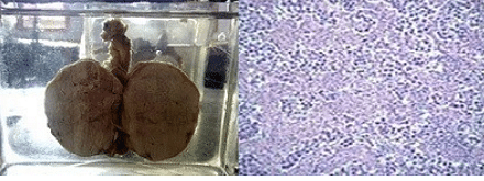
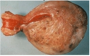
Step 1: Read the histology in the picture as a testicular germ cell tumour and look for the discriminating feature. Step 2: Seminoma arises from the germinal epithelium of the seminiferous tubules and is highly chemo- and radio-sensitive, giving cure rates over $95\%$ when caught early. Step 3: Its hallmark microscopy is monotonous large cells with clear glycogen-rich cytoplasm arranged in nests, partitioned by fibrous bands packed with lymphocytes. Step 4: Teratoma would show multiple tissue types and non-seminoma a mixture of elements, so the clean lymphocyte-rimmed pattern identifies the lesion as seminoma.\[\boxed{\text{Seminoma}}\]